This lab manual, utilizing Marieb/Hoehn’s artwork, enhances understanding of human body structure and function, adaptable to various A&P textbooks.
Specifically, the Cat Version (12th Edition, ISBN 0321971353/978-0321971357) provides a practical, hands-on learning experience for students.
Purpose of the Lab Manual
The primary purpose of this Human Anatomy & Physiology Laboratory Manual, Cat Version, is to supplement and enhance lecture material through practical, hands-on experience. It’s designed to solidify understanding of anatomical structures and physiological processes by allowing students to directly observe and dissect a cat, providing a tangible learning experience.
This manual aims to develop essential laboratory skills, including proper dissection techniques, accurate anatomical identification, and thoughtful observation. Furthermore, it encourages students to apply theoretical knowledge gained in lectures to real-world anatomical examples. The exercises are structured to guide students toward a deeper comprehension of how structure dictates function within the body.
Ultimately, the lab manual serves as a crucial tool for reinforcing concepts and preparing students for further study in the biological sciences, utilizing artwork from established texts like Marieb/Hoehn’s.
Relationship to Human Anatomy and Physiology
While utilizing the cat as a model, this Human Anatomy & Physiology Laboratory Manual, Cat Version, directly correlates to human anatomy and physiology; The cat’s anatomical structures, though differing in some specifics, share fundamental similarities with those of humans, providing a valuable comparative framework for learning.
Students will discover how homologous structures function across species, strengthening their understanding of core physiological principles. Dissection and study of the cat’s systems – skeletal, muscular, nervous, digestive, cardiovascular, and respiratory – illuminate corresponding human systems.
This comparative approach fosters a deeper appreciation for the evolutionary relationships between species and reinforces the applicability of anatomical and physiological knowledge to human health and disease. The manual, adaptable to various A&P texts, ensures a cohesive learning experience.
Overview of the Cat as a Model Organism
The domestic cat serves as an excellent model organism in anatomy and physiology labs due to several key characteristics. Its internal anatomy closely resembles that of humans, making it a valuable tool for understanding human biological systems. The size of the cat facilitates dissection and detailed observation of organs and tissues.
Furthermore, cats exhibit a relatively complete organ system development, mirroring human complexity. This Cat Version of the laboratory manual leverages these advantages, offering students a hands-on experience that bridges the gap between theoretical knowledge and practical application.
Available alongside Main and Fetal Pig versions, the cat model provides a unique learning opportunity, enhancing comprehension of anatomical relationships and physiological functions applicable to human biology.
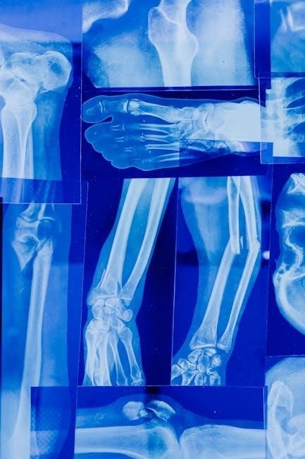
Skeletal System
This section details the cat’s skeletal anatomy, encompassing both axial (skull, vertebral column) and appendicular (limbs) structures, for comparative study.
Cat Skeletal Anatomy: Overview
The feline skeletal system serves as an excellent model for understanding vertebrate anatomy, offering significant parallels to the human skeletal framework. This lab manual’s cat version facilitates comparative analysis, highlighting both similarities and differences in bone structure and articulation.
Students will explore the cat’s skeletal components, including the axial skeleton – comprised of the skull, vertebral column, and rib cage – providing essential support and protection for vital organs. Furthermore, the appendicular skeleton, encompassing the forelimbs and hindlimbs, will be examined in detail, focusing on their role in locomotion and manipulation.
Detailed observation of the cat skeleton allows for a practical understanding of bone classifications, joint types, and the overall biomechanics of movement. This hands-on approach reinforces theoretical knowledge gained from accompanying anatomy and physiology textbooks, enhancing comprehension of human skeletal anatomy as well.
Axial Skeleton – Cat
The axial skeleton of the cat, forming the central support structure, comprises the skull, vertebral column, and rib cage. This section of the lab manual focuses on detailed examination of these components, enabling students to appreciate their protective and supportive functions.
The feline skull exhibits specific features adapted for its predatory lifestyle, including large orbits for binocular vision and a powerful jaw for capturing prey. The vertebral column, divided into cervical, thoracic, lumbar, sacral, and caudal regions, provides flexibility and supports the body’s weight.
Ribs articulate with the thoracic vertebrae, forming the rib cage which safeguards vital organs like the heart and lungs. Careful dissection and observation will reveal the intricate relationships between these bony structures, providing a solid foundation for understanding the axial skeleton’s role in both feline and human anatomy.
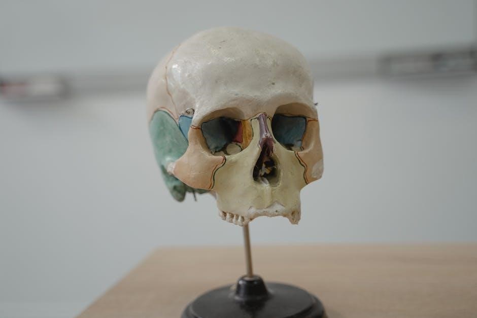
Skull – Cat Specific Features
The cat skull, when compared to the human skull, displays several key adaptations reflecting its carnivorous nature and lifestyle. Notably, cats possess larger orbits – the bony sockets housing the eyes – facilitating enhanced binocular vision crucial for accurate depth perception during hunting.
A shortened facial region and a more pronounced sagittal crest, a bony ridge along the midline of the skull, provide attachment points for powerful jaw muscles. These muscles enable a strong bite force necessary for subduing prey. The feline skull also features a complete bony palate, separating the nasal and oral cavities, allowing for simultaneous breathing and eating.
Students will observe these distinct features during dissection, comparing and contrasting them with human skull anatomy to understand the evolutionary pressures shaping skeletal morphology.
Vertebral Column – Cat
The cat vertebral column, like that of humans, is divided into five regions: cervical, thoracic, lumbar, sacral, and caudal (tail). However, key differences exist reflecting feline locomotion and flexibility. Cats possess a greater number of vertebrae – typically 52-53 – compared to the human count of 33.
The cervical region exhibits increased flexibility, enabling a wide range of head movements. The thoracic vertebrae articulate with the ribs, providing protection for vital organs. The lumbar region supports the abdomen, while the sacral vertebrae fuse to form the sacrum, connecting to the pelvic girdle;
Finally, the caudal vertebrae form the tail, aiding in balance and communication. Students will identify these regions and observe the intervertebral discs, crucial for shock absorption and spinal flexibility.
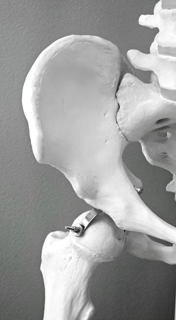
Appendicular Skeleton – Cat
The appendicular skeleton of the cat, encompassing the limbs and their girdles, demonstrates adaptations for quadrupedal locomotion and agility. This section focuses on comparative anatomy, highlighting similarities and differences between feline and human skeletal structures. The pectoral and pelvic girdles provide attachment points for the forelimbs and hindlimbs, respectively.
The forelimbs, including the scapula, humerus, radius, ulna, carpals, metacarpals, and phalanges, are designed for weight-bearing and manipulation. Similarly, the hindlimbs – comprising the pelvis, femur, tibia, fibula, tarsals, metatarsals, and phalanges – facilitate powerful propulsion.
Students will dissect and identify these bones, noting the unique features that contribute to the cat’s remarkable climbing and hunting abilities, furthering understanding of skeletal function.
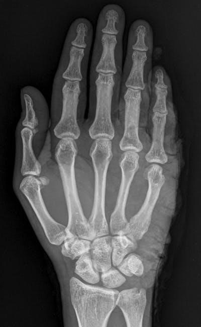
Forelimb – Cat Anatomy
The feline forelimb exhibits a skeletal structure adapted for flexibility and precise movement, crucial for predation and locomotion; Dissection reveals the scapula, providing attachment for muscles, and the humerus, forming the upper arm. The radius and ulna constitute the forearm, enabling rotation.
Carpal bones, analogous to the human wrist, connect to the metacarpals, forming the palm. Finally, the phalanges – the digits – are equipped with retractable claws, essential for grasping and climbing. Comparative analysis with the human forelimb highlights differences in bone proportions and joint mobility.
Students will identify these structures, observing ligament attachments and muscle origins, gaining insight into the biomechanics of feline movement and skeletal adaptations.
Hindlimb – Cat Anatomy
The cat’s hindlimb demonstrates powerful musculature and skeletal adaptations for jumping, running, and maintaining balance. The pelvic girdle, formed by the ilium, ischium, and pubis, provides a stable base for locomotion. The femur, the longest bone in the hindlimb, articulates with the acetabulum of the pelvis.
Distally, the tibia and fibula comprise the lower leg, connecting to the tarsals – equivalent to the human ankle. Metatarsals form the foot, and phalanges constitute the toes, equipped with non-retractable claws for traction. Careful dissection reveals key muscle attachments and ligamentous structures.
Comparative study with the human hindlimb emphasizes differences in limb proportions and the importance of the hindlimb in feline locomotion and agility.
Muscular System
This section explores cat muscle tissue types – skeletal, smooth, and cardiac – and details major muscle groups, aiding comprehension of feline movement and function.
Cat Muscle Tissue Types
Feline muscular systems, like those of humans, comprise three primary tissue types: skeletal, smooth, and cardiac muscle. Skeletal muscle, responsible for voluntary movements, attaches to bones via tendons, exhibiting a striated appearance under microscopy due to organized contractile proteins. This allows for precise control of locomotion and posture.
Smooth muscle, found in the walls of internal organs like the digestive tract and blood vessels, facilitates involuntary functions such as peristalsis and vasoconstriction. Its non-striated structure reflects a different contractile mechanism. Cardiac muscle, exclusive to the heart, possesses characteristics of both skeletal and smooth muscle, exhibiting striations and involuntary control, ensuring rhythmic and efficient heart contractions.
Understanding these distinctions is crucial, as studying cat muscle tissues provides a comparative basis for comprehending human muscular physiology and pathology, enhancing overall anatomical knowledge.
Major Muscle Groups – Cat
Cat musculature, while adapted for feline locomotion, shares fundamental similarities with human muscle groups. Key areas include muscles of the head and neck, facilitating facial expressions and head movement; trunk muscles, supporting posture and enabling body flexion/extension; and limb muscles, powering locomotion.
Forelimb muscles, such as the biceps brachii and triceps brachii, control elbow flexion and extension, mirroring human arm function. Hindlimb muscles, including the quadriceps femoris and hamstrings, drive leg movement. The abdominal muscles provide core stability, while back muscles support the vertebral column.
Studying these groups in cats offers a tangible model for understanding human muscle organization and function, aiding in comparative anatomy and physiological principles.
Muscles of the Head and Neck – Cat
Feline head and neck muscles demonstrate remarkable dexterity, crucial for predation and sensory perception. Muscles like the masseter and temporalis facilitate powerful jaw closure for efficient chewing, while the orbicularis oculi control eyelid movement and blinking. The platysma, a thin sheet of muscle, contributes to facial expression.
Neck muscles, including the sternocleidomastoid and trapezius, enable head rotation, flexion, and extension, mirroring human anatomy. These muscles support the head’s weight and coordinate movements with the vertebral column. Observing these structures in the cat provides a comparative model for understanding human head and neck musculature.
Detailed dissection reveals functional parallels, enhancing comprehension of muscle origins, insertions, and actions.
Muscles of the Trunk – Cat
The feline trunk musculature exhibits a complex arrangement supporting locomotion, posture, and internal organ protection. Key muscles include the external and internal abdominal obliques, rectus abdominis, and transversus abdominis, analogous to human core muscles. These contribute to body wall stability and assist in respiration and defecation.
Back muscles, such as the longissimus dorsi and iliocostalis, extend and laterally flex the vertebral column, enabling agile movements. The diaphragm, crucial for breathing, separates the thoracic and abdominal cavities. Studying these muscles in the cat offers a valuable comparative perspective.
Dissection highlights muscle attachments and functions, reinforcing understanding of mammalian trunk anatomy.
Nervous System
The cat nervous system, explored in this lab, provides a comparative model for understanding human neurological structures, including the brain, spinal cord, and peripheral nerves.
Cat Brain Anatomy
The feline brain, while differing in absolute size from the human brain, exhibits remarkably similar organizational features, making it an excellent model for neurological study.
Key structures, such as the cerebrum, cerebellum, and brainstem, are readily identifiable in preserved specimens, allowing students to trace pathways and correlate structure with function.
This lab focuses on identifying major lobes of the cerebrum, understanding the role of the cerebellum in motor coordination, and recognizing the vital functions controlled by the brainstem – respiration and heart rate.
Dissection exercises and accompanying diagrams facilitate comprehension of gyri, sulci, and the overall architecture of the cat brain, providing a foundational understanding applicable to human neuroanatomy.
Comparative analysis highlights both similarities and differences, reinforcing the principles of evolutionary biology and the conserved nature of essential brain structures across mammalian species.
Spinal Cord and Spinal Nerves – Cat
The cat spinal cord, protected within the vertebral column, serves as a crucial conduit for transmitting signals between the brain and the peripheral nervous system, mirroring human anatomy.
Lab exercises emphasize identifying the dorsal and ventral horns, the central canal, and the gray and white matter organization within a cross-sectional view of the cord.
Students will trace the emergence of spinal nerves through the intervertebral foramina, understanding their role in both sensory input and motor output to specific body regions.
Review sheet questions, potentially assigned via Mastering A&P, reinforce comprehension of spinal cord anatomy and the functional significance of different nerve pathways.
This practical experience builds a strong foundation for understanding neurological reflexes and the impact of spinal cord injuries, concepts directly transferable to human physiology.
Peripheral Nervous System – Cat
The cat’s peripheral nervous system (PNS), analogous to that of humans, comprises the cranial and spinal nerves extending outward from the central nervous system.
Lab investigations focus on identifying major peripheral nerves in the cat, observing their branching patterns, and correlating them with specific muscle groups and sensory regions.
Students will analyze the composition of nerves – sensory and motor fibers – and understand how they contribute to reflex arcs and voluntary movements.
Dissection exercises allow for visualization of nerve plexuses, such as the brachial plexus, and their role in innervating the limbs.
This hands-on approach reinforces understanding of how the PNS connects the CNS to the body, enabling sensory perception and coordinated motor responses, mirroring human neurological function.

Digestive System
The cat digestive system, explored in this lab, provides a comparative model for understanding human digestive anatomy and physiological processes, aiding comprehension.
Cat Digestive Tract Anatomy
The feline digestive tract, detailed within this laboratory manual, offers a valuable comparative perspective to human anatomy and physiology. Dissection and observation reveal key structures, beginning with the oral cavity – teeth adapted for a carnivorous diet, and a muscular tongue facilitating food manipulation.
The esophagus transports ingested material to the stomach, a muscular sac responsible for initial food breakdown through mechanical mixing and enzymatic digestion. Further along the tract, the small intestine – comprised of the duodenum, jejunum, and ileum – is where the majority of nutrient absorption occurs, aided by a large surface area created by villi and microvilli.
Finally, the large intestine, including the cecum, colon, and rectum, primarily functions in water absorption and waste compaction, culminating in fecal elimination. This practical exploration enhances understanding of homologous structures and functional parallels within the human digestive system.
Organs of Digestion – Cat
Detailed examination of feline digestive organs within this lab manual provides crucial insights into comparative anatomy. The stomach, a J-shaped organ, exhibits distinct regions – cardia, fundus, body, and pylorus – responsible for storing and initiating protein digestion via gastric juices. Moving distally, the small intestine’s duodenum receives bile from the liver and pancreatic enzymes, vital for nutrient breakdown.
The jejunum and ileum continue this process, maximizing absorption through their extensive villi. The large intestine, featuring a relatively small cecum in cats, concentrates undigested material. The liver, pancreas, and gallbladder, though accessory organs, are essential for digestion, producing and secreting vital substances.
Understanding these feline structures illuminates corresponding human organ functions, reinforcing core anatomical and physiological principles.
Stomach – Cat
The feline stomach, a J-shaped organ, is a key focus within this lab manual’s digestive system section. Dissection reveals distinct anatomical regions: the cardia, where the esophagus enters; the fundus, a dome-shaped expansion; the body, the stomach’s main central portion; and the pylorus, leading to the duodenum. Histological examination highlights the stomach’s layered structure – mucosa, submucosa, muscularis externa, and serosa.
The mucosa contains gastric pits and glands, secreting hydrochloric acid and pepsinogen, initiating protein digestion. The muscularis externa’s contractions facilitate mechanical breakdown and mixing of food. Observing these features in the cat stomach provides a tangible understanding of mammalian digestive processes, directly correlating to human anatomy and physiology.
This comparative approach enhances learning.
Intestines – Cat
The cat’s intestinal system, explored in this lab manual, comprises the small and large intestines, crucial for nutrient absorption and waste processing. The small intestine – duodenum, jejunum, and ileum – exhibits features like plicae circulares and villi, maximizing surface area for absorption. Dissection reveals the mesenteric vessels supplying these regions.
The large intestine, including the cecum, colon, and rectum, primarily reabsorbs water and forms feces. Examining the intestinal walls microscopically demonstrates the distinct histological structures adapted for their respective functions. Comparing the cat’s intestinal anatomy to the human system reinforces understanding of mammalian digestive physiology.
This hands-on experience solidifies key concepts.
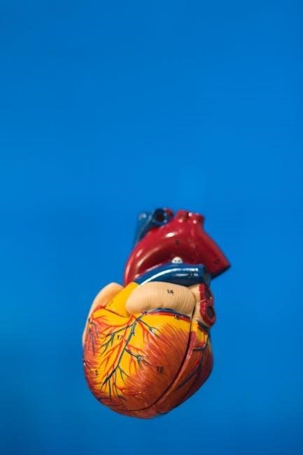
Cardiovascular System
This section details the cat’s heart anatomy and blood vessel network, providing a comparative understanding relevant to human cardiovascular physiology within the lab manual.
Cat Heart Anatomy
The feline heart, explored within this lab manual’s cardiovascular system section, presents a valuable model for understanding mammalian cardiac structure and function, closely mirroring human anatomy. Dissections reveal the four chambers – two atria and two ventricles – essential for efficient blood circulation.
Students will identify key features like the aorta, vena cava, pulmonary artery, and pulmonary veins, tracing blood flow pathways. The manual guides observation of the pericardium, the protective sac surrounding the heart, and the myocardium, the muscular heart wall.
Comparative analysis highlights similarities and differences between the cat and human heart, reinforcing concepts of cardiac physiology. This hands-on experience, utilizing the 12th Edition’s resources (ISBN 0321971353/978-0321971357), solidifies comprehension of circulatory mechanisms.
Blood Vessels – Cat
This section of the lab manual details the feline circulatory network, providing a practical understanding of blood vessel structure and function, directly applicable to human physiology. Students will dissect and identify major arteries – aorta, carotid, femoral – and veins – vena cava, jugular, saphenous – tracing blood’s journey.
Capillaries, the microscopic exchange vessels, are discussed in relation to tissue perfusion. The manual guides observation of vessel wall layers – tunica intima, media, and adventitia – and their role in maintaining blood pressure and flow.
Comparative study emphasizes the similarities between feline and human vascular systems, reinforcing core concepts. Utilizing the 12th Edition (ISBN 0321971353/978-0321971357), students gain a comprehensive grasp of cardiovascular anatomy and its vital role in sustaining life.

Respiratory System
This lab manual section explores feline respiratory organs, aiding comprehension of gas exchange and ventilation, mirroring human anatomical and physiological principles.
Cat Respiratory Organs
The feline respiratory system, detailed within this lab manual, provides a comparative model for understanding human respiration. Dissections and observations focus on the nasal cavity, pharynx, larynx, trachea, bronchi, and lungs – structures homologous to those found in humans.
Students will identify key anatomical features, tracing the pathway of air and examining the microscopic structure of lung tissues. This hands-on approach reinforces understanding of gas exchange mechanisms, including the role of alveoli. The manual, often accompanying texts like Amerman’s Human Anatomy & Physiology, facilitates applying concepts learned in lecture to practical laboratory settings.
Comparative analysis highlights similarities and differences between feline and human respiratory systems, deepening comprehension of functional adaptations. The cat model effectively illustrates respiratory physiology, preparing students for advanced studies in human anatomy and physiology.